Chapter 3. Chromosomal Composition and Nomenclature
Chromosomes And Chromatin
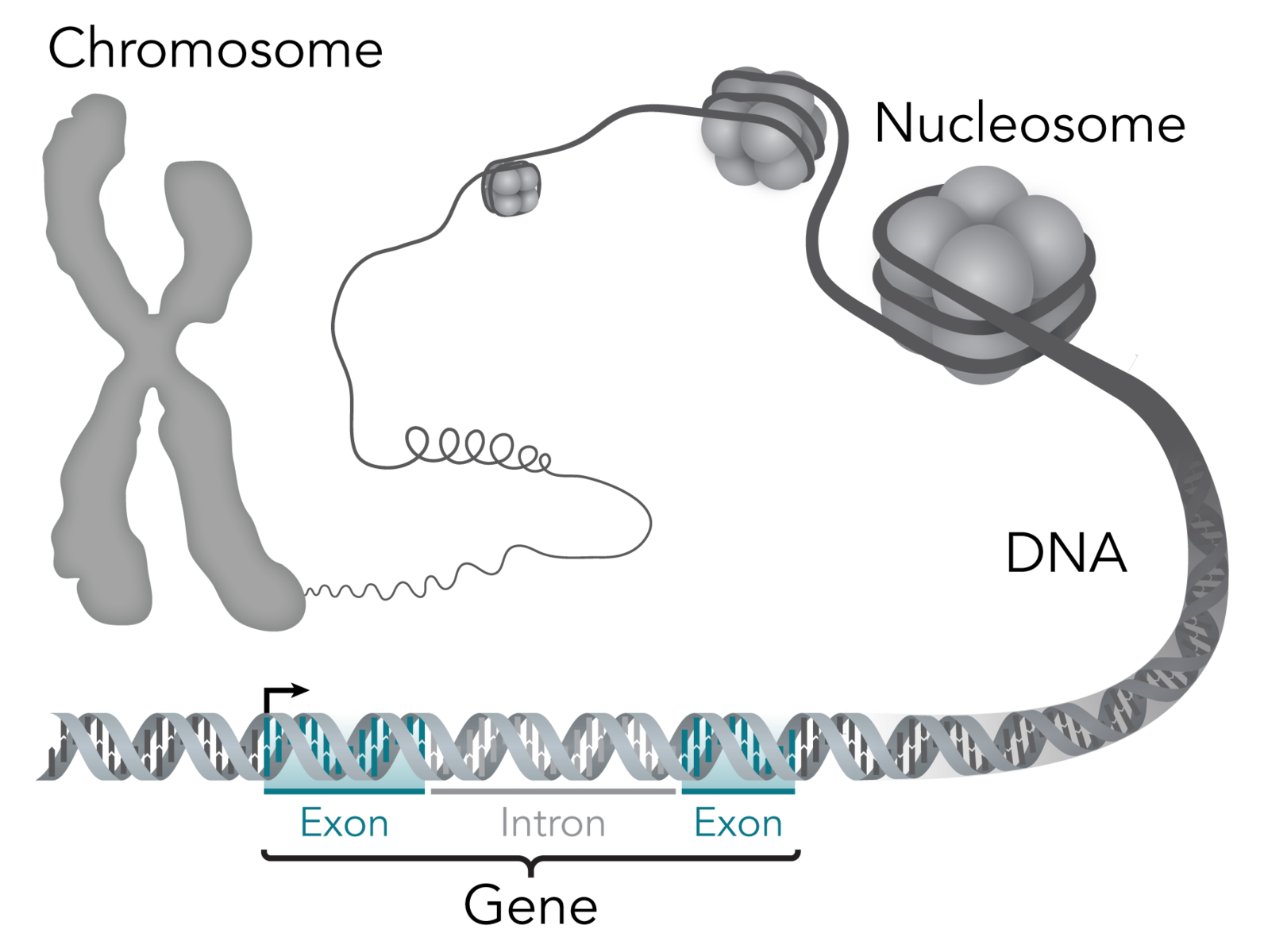
DNA in human cells is organized into chromosomes. All eukaryotic organisms have chromosomes, which are discrete pieces of double-stranded DNA, wrapped around nuclear proteins called histones. The arrangement of DNA and nuclear proteins is referred to as chromatin. The number of chromosomes varies from species to species. The diploid number of human chromosomes is 46.
Just before cell division (and after DNA synthesis), the chromatin condenses into individual chromosomes. The dividing chromosomes appear as two chromatids. The chromatids appear to be made of coiled loops of 20-30 nm-thick chromatin fibers.
The structural unit of chromatin is the nucleosome.
Each nucleosome consists of eight units of nuclear proteins called histones (two each of histones H2A, H2B, H3, and H4) associated with 146 nucleotide pairs of DNA and a stretch of linker DNA of varying length. The diameter of the nucleosome “bead,” or core particle, is about 10 nm. Histone H1 is thought to bind to the linker DNA and facilitate the packing of nucleosomes into 30-nm fibers.
Chromosome nomenclature
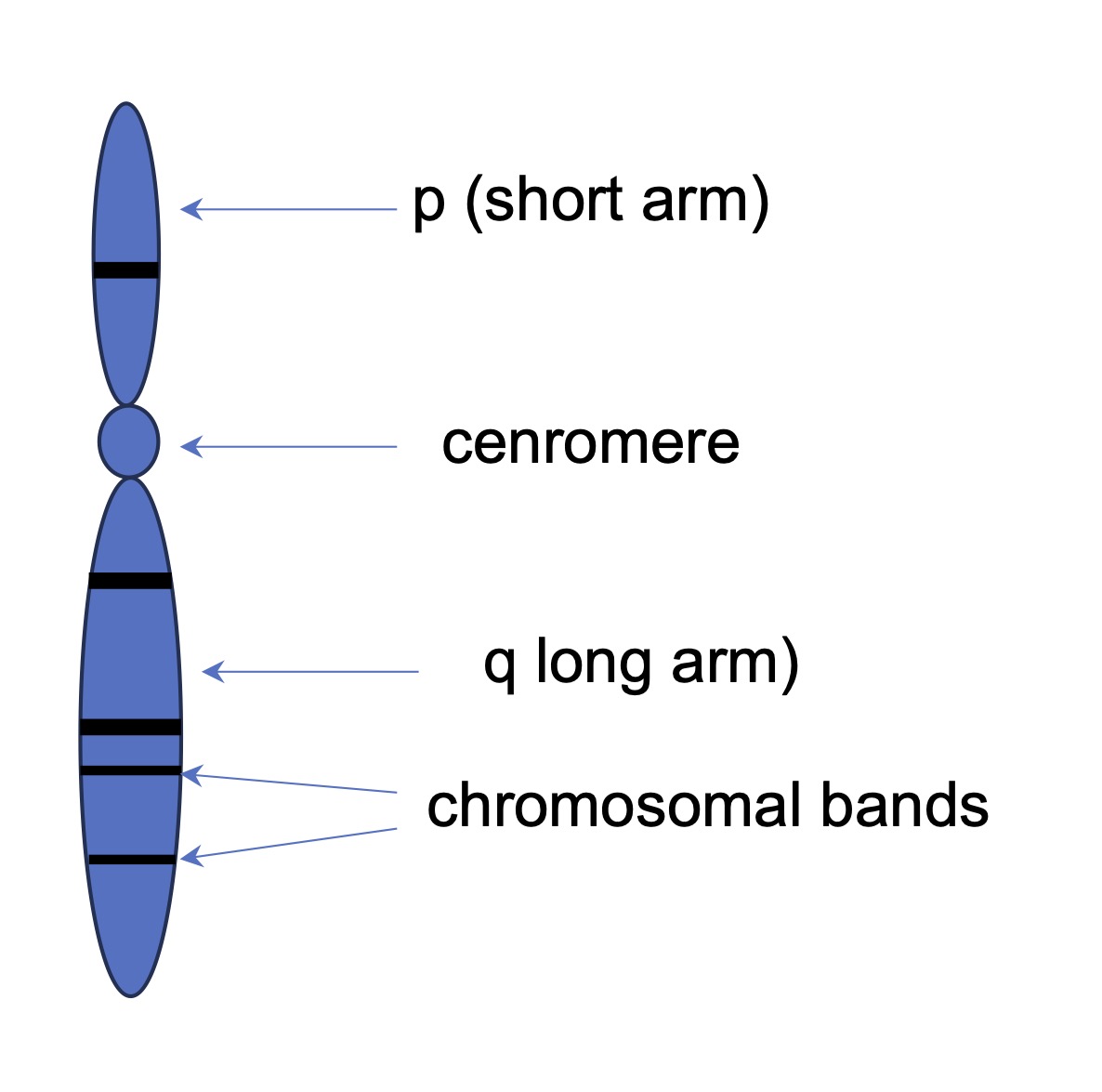
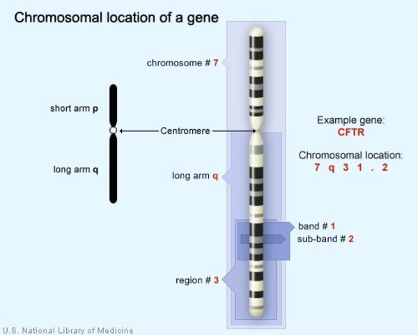
In human and other genomes, chromosomes are broken down into arms, regions, and bands.
Chromosome arms are defined with respect to the kinetochore (centromere). A centromere is a region of DNA found along the length of a chromosome where two identical sister chromatids come in contact. It is involved in cell division as the point of mitotic spindle attachment. Centromeres function in sister chromatid adhesion and pairing of homologous chromosomes during meiosis. During mitotic and meiotic divisions, a protein structure called kinetochore is formed on top of the centromeres. The kinetochores are the sites where the spindle fibers attach during the cell division to pull the chromosomes apart.
The centromere usually contains specific tandem repetitive DNA sequences, which are often called “satellite DNA”. At the end of chromosomes, DNA sequences called telomeres are composed of the same type repetitive sequences as well. The centromeres are, together with telomeres and origins of replication, one of the essential parts of any eukaryotic chromosome.
Although the terms “kinetochore” and “centromere” are used interchangeably in chromosome nomenclature, it is probably more correct to refer to kinetochore, since we don’t really see the centromere, which is simply a DNA sequence. Rather, we see the proteinaceous kinetochore, and the distinct heterochromatic staining that occurs in the vicinity of the centromere.
On either side of the centromere there are ‘arms’ that extend to terminal regions, known as telomeres. The short arm of a chromosome is designated ‘p’ (p = “petite”), while the long arm is referred to as ‘q.’ The band nomenclature refers
When chromosomes are physically stained with a Giemsa dye, dark-colored horizontal lines b become visible on both chromosomal arms. This G-banding is most visible on chromosomes during the metaphase stage of cell division. Bands are numbered outward from the centromere with the largest values near the telomeres.
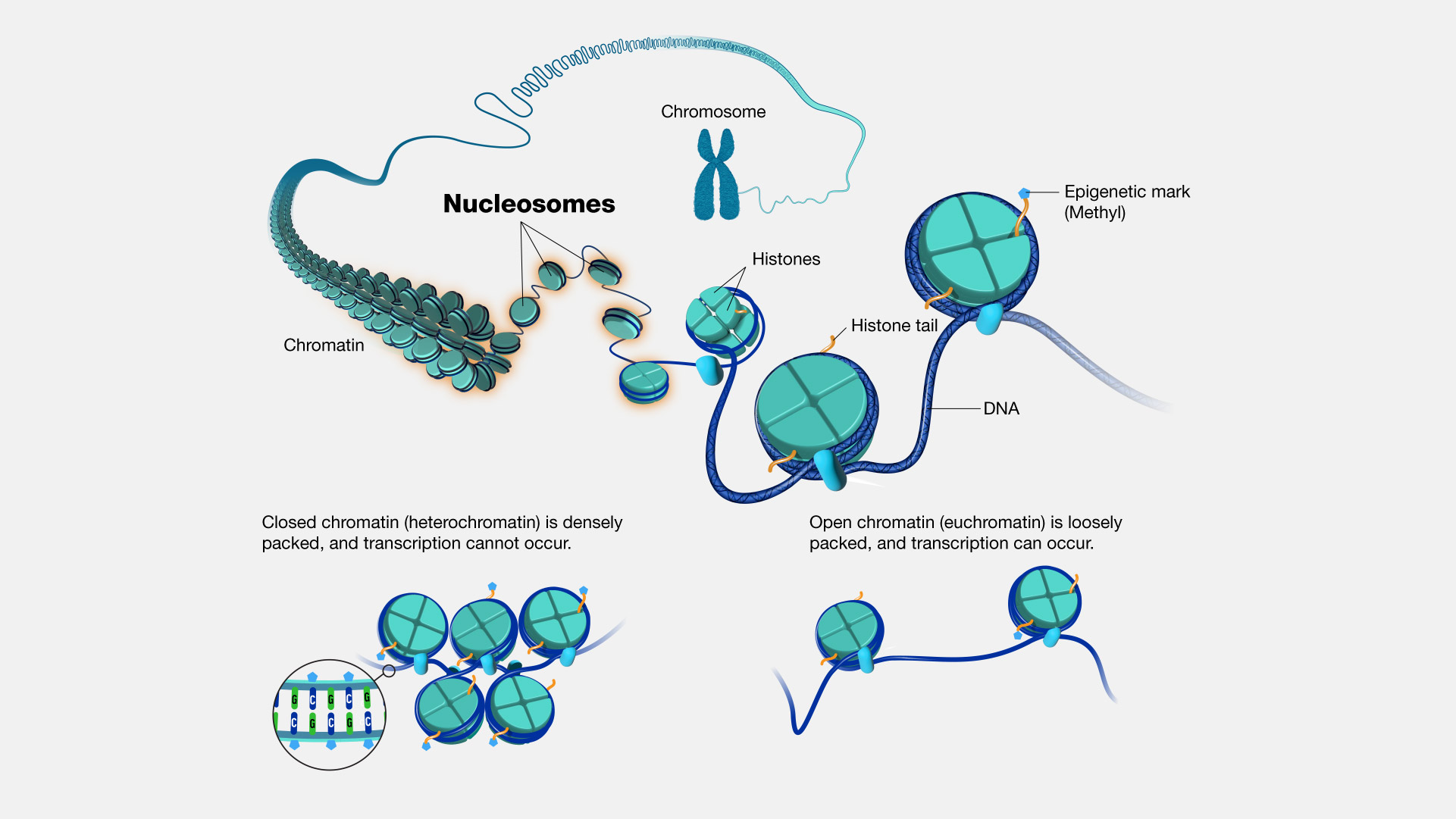
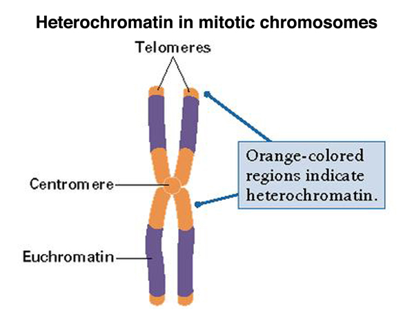
Heterochromatin and euchromatin
Heterochromatin is a tightly packed form of DNA, whereas euchromatin is a lightly packed form. Heterochromatin is found in genetically inactive regions, such as the centromeric and telomeric regions of chromosomes. When stained with dyes used in chromosomal studies such as polychrome methylene blue-eosin Y (Giemsa stain), heterochromatin stains darker (more intensely) than euchromatin. Euchromatic regions usually display a higher amount of genetic activity, and the lighter packing allows access to the information encoded into the DNA of euchromatic regions.
Types of chromosomes
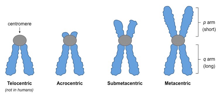
There are three types of chromosomes in humans, based upon the position of the centromere.
Metacentric: In this type of chromosome the arms are of equal length. Human chromosomes 1, 3, 16, 19, 20 are metacentric.
Submetacentric: In this type of chromosome one arm is slightly longer than the other. Human chromosomes 2, 4, 5, 6-12, 17, 18, X are submetacentric.
Acrocentric: In this type of chromosome one arm is significantly longer than the other.
A small round or rod-shaped structure called satellite, attached by a very thin thread is variably present on the short arm of acrocenrtic chromosomes. There are five acrocentirc chromosomes with satellites in the human genome: 13-15, 21, and 22. Chromosome Y is considered acrocentric as well but has no satellites.
Banding pattern of human chromosomes
Chromosomes in metaphase can be identified using certain staining techniques. Most conventional cytogenetic analyses depend on the karyotyping of banded metaphase chromosomes.
A band is defined as that part of a chromosome which is clearly distinguishable from its adjacent segments by appearing darker or brighter when stained with a dye. In G-bands (produced by Giemsa dye), the dark regions tend to be heterochromatic, whereas the light regions tend to be euchromatic. Hatched pattern represents regions that vary in staining intensity.
The chromosomal nomenclature starts with the chromosome itself. For example, 15q represents the long arm of chromosome 15. The first digit after the arm indicates the region within the arm. Numbers increase going away from the kinetochore on each arm: 15p1, 15p2, 15p3, or 15q1, 15q2, 15q3, and so on. The next digit indicates specific band within that region. Such as, 15q13 refers to chromosome 15, long arm, region 1, band 3. Digits after decimal points indicate additional bands resolved more recently after the original nomenclature was established in 1971.
Key Takeaways
- DNA in human cells is organized into chromosomes composed of euchromatin and heterochromatin.
- The structural unit of the chromosome is the nucleosome.
- Chromosomal nomenclature takes into account chromosomal arms as well as bands that appear on a stained chromosome.
Chromosomes are DNA molecules in association with proteins that contain genetic information arranged in a linear sequence.
Chromatin is the complex of DNA and proteins that makes up a chromosome.
A tightly packed (and darker stained) form of chromatin usually associated with areas of low or no gene expression (such as centromeres and telomeres). Heterochromatic regions are heavily methylated.
A lightly packed (and lightly stained) form of chromatin that is enriched in genes, and is usually associated with active gene expression.
The type of chromosome in which one arm is slightly longer than the other arm. Human chromosomes 2, 4, 5, 6-12, 17, 18, X are submetacentric.
