6 Ventricular Arrhythmias

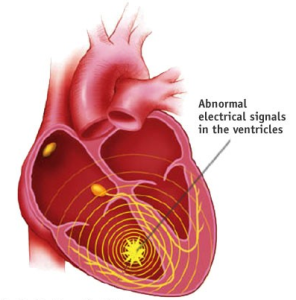
Within the ventricles, the SA node (sinus signal) coming through the AV node is conducted rapidly along the bundle branches, outward through the purkinje fibers and across the entire ventricle rapidly. This results in a simultaneous contraction of the left and right ventricle, and ejection of blood. Premature ventricular contractions discussed previously, often occur as a result in increased excitation of the Purkinje fibers, resulting in an isolated PVC. However, more extensive ventricular arrhythmias are possible due to both cardiac and non-cardiac conditions, resulting in completely new rhythms, some of which can be fatal if not corrected immediately.
Ventricular Bigeminy, Trigeminy – atria and ventricle sharing the pacing
In ventricular bigeminy, we see a sinus beat, followed by a PVC (ventricular beat). Then there is a pause, followed by another sinus beat-then ventricular beat pairing. The rhythm can be continuous, or intermittent, or can be of cardiac origin (myocardial injury or ischemia) and non-cardiac origin (stimulants, alcohol, hypokalemia, anxiety). The nature of the sinus to ventricular pacing can vary from 1:1 (bigeminy), 2:1 (trigeminy) 3:1 (quadrigeminy) and so on.


Ventricular Tachycardia
In many cases, ventricular tachycardia is a result of heart disease and scarring present in the myocardium. The scarring creates the possibility of a reentrant, fast paced tachycardia originating in the ventricle. A well times PVC may be the trigger initiate ventricular tachycardia (“Vtach”). Vtach may be referred to as non-sustained (lasts under 30 seconds) or sustained. If the rate is high, the patient may feel dizzy, may have a pallor complexion, and possibly not show a pulse due to the fast rate (reduced diastolic filling time). Rates are greater than 100 and could approach 300bpm. Since the signal is originating in the ventricle, there are no P waves present. Since the signal is also occurring outside of the bundle branch conduction system, the QRS complex is wide.
Danger! This rhythm can lead to sudden cardiac death. Treatments for Vtach include:
- – Cardioversion, similar to atrial fibrillation
- – In no-pulse Vtach, defibrillation may be indicated
- – Longer term treatment may include medications to suppress Vtach reoccurrence.

Watch this animation of ventricular tachycardia. Courtesy the American Heart Association. Be sure to select ventricular tachycardia from the menu on the lower right of image.


Polymorphic Ventricular Tachycardia- « Torsades de Pointes »
One unique version of wide complex ventricular tachycardia is often seen in patients with long QT intervals. Long QT can be inherited or induced as a side effect of some drugs. This form of Vtach shows changes in the morphology of the ventricular beats, often oscillating from tall to short, like twisting a slinky. The term “Torsades de pointes” is French for “twisting of peaks”.

Sudden Cardiac Death: Ventricular Fibrillation
With ventricular fibrillation, there is a loss of all organized contraction in the ventricles. Instead, there is disorder electrical activity across both ventricles, resulting in the ventricles “quivering” with a lack of any contraction. Cardiac output drops to nothing, the patient is unconscious, and death will occur if not treated with defibrillation as soon as possible. CPR can serve as a brief bridge to extend the time before defibrillation occurs. The ECG characteristics of Ventricular fibrillation (“V Fib”) is disordered electrical waves of with large (course Vfib) or small magnitude (fine Vfib). Patients that survive conversion out of Vfib are often fitted with implantable defibrillators that can sense the onset of vfib and apply an immediate shock to revive the patient.

Watch this animation of ventricular fibrillation. Be sure to select ventricular fibrillation from the menu on the lower right of the image.


Lack of Systolic Contraction: Cardiac Standstill or Asystole


Selected Sources
Goldberger’s Clinical Electrocardiography: A simplified approach 10th ed. Goldberger A., Goldberger, Z, Shvilkin A. Elsevier pub. ISBN-10 0323824757 2023.
Clinical Exercise Electrocardiography – Levine S., Coyne B., Colvin L. Jones & Bartlett Learning Pub. ISBN-10 1284034208 2015.
https://hqmeded.ecg.blogspot.com
Video links used in Chapter 6
Animation of ventricular tachycardia (select vtach from menu on lower right of image): https://watchlearnlive.heart.org/?moduleSelect=arrhyt
Animation of ventricular fibrillation (Be sure to select ventricular fibrillation from the menu on the lower right of the image): https://watchlearnlive.heart.org/?moduleSelect=arrhyt
A timed defibrillation procedure where the depolarization shock is synchronized with the ventricular systole to prevent inducing dangerous arrhythmias. It is used in cases of atrial fibrillation, flutter and SVT. Chemical cardioversion is also possible with medications such as flecainide, amiodarone or propafenone.
A medical treatment for life threatening arrhythmias, such as ventricular fibrillation, where cardiac cells randomly fire, resulting in no coordinated ventricular contraction. Defibrillation sends an electric shock through the heart, depolarizing all cells at once, with the hope that the SA node resumes its role as the normal pacemaker. Defibrillation can be done externally to the chest, and internal defibrillators can be implanted as part of a pacemaker.
